43 label the photomicrogram of the trachea.
Solved Label the photomicrogram of the trachea. Cilia Lamina | Chegg.com Anatomy and Physiology questions and answers. Label the photomicrogram of the trachea. Cilia Lamina propria Submucosa Cilia Basement membrane Submucosa Epithelium Basement membrane Lamina propria Epithellum. Question: Label the photomicrogram of the trachea. Cilia Lamina propria Submucosa Cilia Basement membrane Submucosa Epithelium Basement ... Free Automated Malware Analysis Service - powered by Falcon Sandbox ... Submit malware for free analysis with Falcon Sandbox and Hybrid Analysis technology. Hybrid Analysis develops and licenses analysis tools to fight malware.
A & P lab test 4 Flashcards | Quizlet Label the photomicrogram of the trachea. Correctly label the following anatomical features of the lower respiratory tract. ... Drag each label into the appropriate position in order to identify whether the term or item is involved with chemical or mechanical digestion.
Label the photomicrogram of the trachea.
The Bronchi: Anatomy, Function, and Treatment - Verywell Health The bronchi are the airways that lead from the trachea into the lungs and then branch off into progressively smaller structures until they reach the alveoli, the tiny sacs that allow for the exchange of oxygen and carbon dioxide in the lungs. While the bronchi function primarily as passageways for air, they also play a role in immune function. A&P 139 Chapter 19 Flashcards | Quizlet Label the photomicrogram of the lung. Label the photomicrogram of the trachea. Cricoid. Which of these laryngeal cartilages is single? Label these structures of the upper respiratory system. tidal volume. The volume of air that enters (or … Lab 2: Microscopy and the Study of Tissues - UW-La Crosse This slide showing a cross section of the mammalian trachea (wind pipe) contains examples of several different kinds of tissues. In addition to the pseudostratified columnar epithelium lining the trachea and hyaline cartilage, also seen on this slide is an extensive area of adipose tissue, which is specialized for fat storage.
Label the photomicrogram of the trachea.. Label The Photomicrograph Of The Lung : 4 Chloro Dl Phenylalanine ... The lower respiratory system., put the following layers of the trachea in order from superficial to deep., label the structures of the upper respiratory . Learn what you need to know about lung cancer. My image 1 my image 2. After completion of this video you will be able to: Label the photomicrogram of the lung segmental branch of pulmonary a. The Trachea (Human Anatomy): Picture, Function, Conditions, and More The trachea, commonly known as the windpipe, is a tube about 4 inches long and less than an inch in diameter in most people. The trachea begins just under the larynx (voice box) and runs down ... Histology, Alveolar Cells - StatPearls - NCBI Bookshelf Alveoli represent the major sites of gas exchange. Their presence increases the surface area of the lung to maximize gas exchange, much like villi and microvilli increase the absorptive surface area of the digestive tract. For alveoli to carry out their function efficiently, they must be both a dynamic and stable system. The lung parenchyma must be able to expand and recoil during inspiration ... quizlet.com › 381483065 › a-p-lab-test-4-flash-cardsA & P lab test 4 Flashcards | Quizlet Label the micrograph of the renal corpuscle and surrounding structures using the hints provided. Correctly label the following anatomical parts of a kidney. Classify each of the following parts of the nephron into the correct category based on whether it can only be found in the cortex or if it can be found in the medulla and/or cortex of the ...
Labeled diagram of the lungs/respiratory system. - SERC View Original Image at Full Size. Labeled diagram of the lungs/respiratory system. Image 37789 is a 1125 by 1408 pixel PNG Uploaded: Jan10 14. Last Modified: 2014-01-10 12:15:34 Trinidad State College Home Page Trinidad State is a Hispanic-Serving Institution (HSI) HSI is defined in federal law (the Higher Education Opportunity Act, Title V, 2008) as an accredited, degree-granting, public or private nonprofit institution of higher education with 25% or more total undergraduate Hispanic full-time equivalent (FTE) student enrollment. Medical Speech-Language Pathology: A Desk Reference, Third Edition ... write on label s.o.s. si opus si if necessary ss semis a half stat. statim immediately t.d.s. ter die sumendum to be taken three times daily t.i.d. ter in die three times a day t.i.n. ter in nocte three times at night ut dict ut dictum as directed v.i. vide infra see below via via by way of viz viz namely v.s. vide supra see above a Note: q.i.d ... Anatomy A215 Virtual Microscopy - Indiana University Bloomington Anatomy A215 Virtual Microscopy. Each alveolus is a small air space surrounded by an extensive capillary network. The epithelium which lines the alveoli is an extremely thin simple squamous in close proximity to the. capillary walls. Alveoli make up the major part of the lung.
(Lee Ann C. Golper) Medical Speech-Language Pathol (BookFi) - Scribd [Lee_Ann_C._Golper]_Medical_Speech-Language_Pathol(BookFi).pdf - Free ebook download as PDF File (.pdf), Text File (.txt) or read book online for free. A&P 2 Lab Unit 2 Flashcards | Quizlet Label the photomicrogram of the lung. Identify the cartilaginous anatomical structures shown in the posterior view of the superior portion of the lower respiratory system. Place the following structures with the appropriate anatomical region. Labels maybe be placed in more than one category. ... Label the photomicrogram of the trachea. EOF (Get Answer) - Determine the angle of i, r and q this is reflection of ... Determine the angle of i, r and q this is reflection of light General Structure of Mucosa Label the structures that comprise the respiratory tract mucosa (mucous membrane). ... lung Segmental bronchus Trachea prey="" 11="" of="" 46="" next=""> Trachea histology of respiratory system low power Label the photomicrogram of the trachea.
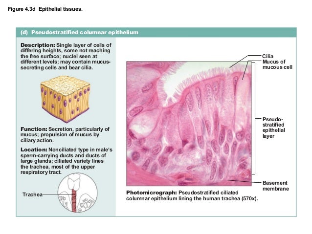
Ex 6a epithelial_tissues
Can you label the lungs? Quiz - PurposeGames.com Printables and Stats. lungs. respiratory system. Labeling the Fetal Skull Bones. 7p Image Quiz. Label the Stomach 9p Image Quiz. Labeling the Cochlea 7p Image Quiz. 8p Image Quiz. Anatomy of the Long Bone 11p Image Quiz. Rib Cage Muscles 2p Image Quiz.

lung histology labeled - bronchiole, alveolar duct, alveoli | lab ...
downloads.chartnettech.com o, o= +, o, o> (- o oI X 2Ð o? Ži 1 o‚ Þ , o@ Ü * ,Z† sS * sU * sM * sP * sc * se * s] * s_ * sg * s[ *.( € * {A * {B * {C * (' * (' *ž {D {E {D {F oG {H ...
The Histology Guide | Respiratory
Histology of trachea and lung - SlideShare 1. HISTOLOGY OF TRACHEA AND LUNG Dr.ushakannan,Asst.professor. 2. RESPIRATORY SYSTEM Conducting Part- responsible for passage of air and conditioning of the inspired air. Examples:nasal cavities,pharynx, trachea, bronchi and their intrapulmonary continuations. Respiratory Part-involved with the exchange of oxygen and carbondioxide between blood ...

Anatomy And Physiology Archive | October 25, 2017 | Chegg.com
BIO208 Lab Practical 2 - 10/6/2019 Lab Practical 2 Home - Course Hero 10/6/2019 Lab Practical 2 Question Label the structure with a "star" symbol beside it. 4 Incorrect Mark 0.00 out of 1.00 Answer: trachea The correct answer is: larynx (based on the document attached to the question as reference)

trachea histology labeled - Google Search | Histology | Pinterest
quizlet.com › 514038744 › ap-139-chapter-19-flash-cardsA&P 139 Chapter 19 Flashcards | Quizlet Label the photomicrogram of the lung. Label the photomicrogram of the trachea. Cricoid. Which of these laryngeal cartilages is single? Label these structures of the upper respiratory system. tidal volume. The volume of air that enters (or leaves) during a single respiratory cycle is the.
Post a Comment for "43 label the photomicrogram of the trachea."