41 label the following photomicrographs by tissue type
ESPIRITU_Activity5.pdf - Activity #5 ANIMAL HISTOLOGY... View ESPIRITU_Activity5.pdf from BIO 24 at De La Salle University. Activity #5 ANIMAL HISTOLOGY Student's Name Course/Section Instructor's Name Date Performed _ Date Submitted I. Label the following Molecular Expressions Photo Gallery: Mitosis - MagLab Mitosis is the mechanism that allows the nuclei of cells to split and provide each daughter cell with a complete set of chromosomes during cellular division. This, coupled with cytokinesis (division of the cytoplasm), occurs in all multicellular plants and animals to permit growth of the organism. In this part of the Photo Gallery, we ...
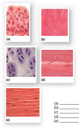
Solved Label the following photomicrographs by tissue type | Chegg.com question: label the following photomicrographs by tissue type and by using the terms provided a. dollagenous fibers, elastic fibers, reticular fibers 2 courtesy of eric wise tissue name (400) b) basement membrane, cilia, nucleus cilia -2 nucleus - basement humbrane courtesy of eric wise tissue name (400x) c. intercalated discs, nucleus, …

Label the following photomicrographs by tissue type
PDF Paper 1 Exam Questions (Live) - Mindset Learn The following table shows an analysis of the nutrients found in 100g portion of breakfast. The breakfast was ... Give two reasons by referring to the photomicrographs. (2) 2.2.2. Label structure 1. (1) 2.2.3. ... The photographs below show various human tissues 3.5. Identify each tissue type labeled A, ... SC 2115 Anatomy and Physiology I - Mass These vary in name depending upon the tissue. 1. Fibroblasts are star-shaped cells responsible for the production of fibers and the matrix. 2. Fibrocytes are mature fibroblasts which maintain connective tissues. 3. Mast cells produce the anticoagulant, heparin, as well as th e irritating vasodilator, histamine. 4. Detection of BrdU-label Retaining Cells in the Lacrimal Gland ... The photomicrographs depicted in Figure 2a-d show that following the 2- and 4-week chase periods, multiple BrdU + cells could be observed in any given lacrimal gland lobule. Figure 2a-g show that the number of BrdU + cells was high after 2 weeks of chase, decreased slightly by the 4 th week and then declined dramatically at 12 weeks.
Label the following photomicrographs by tissue type. PDF Hudson City Schools / Homepage Once everyone is done, complete Qyestion #9 as a group. a. Label the apical surface on each epithelial tissue photomicrograph. b. Label the basal surface on each epithelial tissue photomicrograph. c. Draw a bracket to indicate the location of the epithelial tissue. d. Name the specific epithelial type under both tissue photomicrographs. identify the three phases of mitosis in the following photomicrographs Identify t he four phases of mitosis shown in the following photomicrographs, and select the events from t he key that correctly identify each phase. Explain the role of the structures labelled 2 in the cell division process. Mitosis involves four stages: prophase, metaphase, anaphase, and telophase. 9. | M19-01 photomicrographs of motoneuronal labeling following different ... Download scientific diagram | | M19-01 photomicrographs of motoneuronal labeling following different CAV-2 dosage injections into the (A) medial rectus and (B) lateral rectus muscles. Arrows ... Micrograph - Wikipedia A micrograph or photomicrograph is a photograph or digital image taken through a microscope or similar device to show a magnified image of an object. This is opposed to a macrograph or photomacrograph, an image which is also taken on a microscope but is only slightly magnified, usually less than 10 times.
Label The Photomicrograph Of The Lung : 4 Chloro Dl ... - Blogger Label the photomicrogram of the lung segmental branch of pulmonary a. Relative amounts of glands, cartilage, smooth muscles and connective tissue fibers present in the wall of the tubes. Photomicrographs of bronchioles and pulmonary alveoli of giant anteater (myrmecophaga tridactyla). Learn what you need to know about lung cancer. Chapter 6 Problem 25RQ Solution - Chegg ISBN-13: 9780077676636 ISBN: 0077676637 Authors: Eric Wise Rent | Buy. This is an alternate ISBN. View the primary ISBN for: null null Edition Textbook Solutions. Bi 233 Lab Exercise 36 - Portland Community College Be certain to include a label with the following information: a. Title - What is the drawing of? b. Total Magnification. c. Labels indicating the main items of interest in the drawing (e.g. goblet cells, epithelial cells, and other tissue types, alveoli, etc.) Note: A good resource is "Histology Atlas" on pp. 687 - 697. Tissue Identification Flashcards | Quizlet Explain how each plant tissue has a similar function to the organ or organ system in the human body. (a) dermal tissue and human skin (b) vascular tissue and the circulatory system (c) ground tissue and the skeletal system. Verified answer. BIOLOGY.
Answered: The following are photomicrographs of… | bartleby Label the cell wall, vacuole, and tonoplasts in each photomicrograph and describe these in terms of their shape and color. Question The following are photomicrographs of T. spathacea leaf cells in 0.01 M NaCl and 0.30 M NaCl solutions. CH. 19 Assessment Flashcards | Quizlet Iron is transported in the blood by transferrin. true Organize the steps of the common pathway of blood clotting in the correct sequence from beginning to end. Label the photomicrograph using the hints provided. Match the plasma protein with its description. ALBUMIN: Plays a major role in maintaining colloid osmotic pressure Blood cell histology Flashcards - Quizlet HDonica Terms in this set (4) Place the following terms and descriptions with the appropriate cell that is in the center of each of these histology slides of white blood cells. Label the types of cells in the photomicrograph using the hints provided. Label the photomicrograph using the hints provided. Thyroid and parathyroid label the following organs - Course Hero Label the following on the histology diagram: Thyroid tissue and Parathyroid tissue Follicle Colloid Follicular cells Parafollicular cells Identify the cells that produce the 4 hormones that come from these tissues. placement of the thyroid within the neck histology of the thyroid and parathyroid
Photomicrographs of SEC sections from different treatments: a ...
Act05-Barrento.docx - Activity #5 ANIMAL HISTOLOGY AND... The function of connective tissues involves enveloping muscles such as supplying the body with heat like brown adipose, supporting framework of organs like the bones. Mainly, binding and supporting, protecting, insulating, storing reserve fuel and transporting substances within the body are the major functions of connective tissue. 3.

Brief exposure of neonatal testis cells to EGF or GDNF alters ...
Tissues.docx - McKinley Swif 1/23/2022 Tissues 1. List the... - Course Hero McKinley Swif 1/23/2022 Tissues 1. List the four main tissues of the body. a. Epithelial, connective, muscular, and nervous a. Epithelial , connective , muscular , and nervous 2. What is the name of the noncellular layer that attaches epithelial tissue to other layers? a. Basement membrane a. Basement membrane 3.
Solved b. c. 18. What type of connective tissue is | Chegg.com
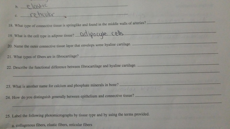
Chapter 4: Tissues Flashcards | Quizlet What type of connective tissue is spring like and found in the middle walls of arteries? ... Label the following photomicrographs by tissue type and by using the terms provided. a. a. elastic fibers, b. collagenous fibers, c. reticular fibers b. a. cilia, b. nucleus, c. basement membrane

Photomicrographs from rat tongue mucosa following 4NQO ...
PRACTICAL 2 -TISSUES Flashcards | Quizlet In connective tissue there are more non-cellular structures present such as fibers or a matrix. Label the following photomicrographs by tissue type and by using the terms provided. a. a. elastic fibers, b. collagenous fibers, c. reticular fibers. b. a. cilia, b. nucleus, c. basement membrane.
Minjuvi® receives EC approval to treat large B-cell lymphoma
LBYZOOL Act.05 Animal Histology and Organology Exercise.pdf... 1. List down fifteen guidelines or guideposts that you wish to follow or you are following in relation to yourself, to other people, to the world and environment, and to God.

Multiple Myelolipomas in Bilateral Posterior Mediastinum ...
Lab 2: Microscopy and the Study of Tissues - UW-La Crosse Epithelium is a type of tissue whose main function is to cover and protect body surfaces but can also form ducts and glands or be specialized for secretion, excretion, absorption and lubrication. Epithelial tissues are classified according to the number of cell layers that make up the tissue and the shape of the cells.

Photomicrograph of a solid ameloblastoma specimen at 400x ...
Solved 25. Label the following photomicrographs by tissue | Chegg.com Question: 25. Label the following photomicrographs by tissue type and by using the terms provicu. a. collagenous fibers, elastic fibers, reticular fibers Courtesy of Eric Wise tissue name (400x) collagenous fibers, elastic fibers, reticular fibers Courtesy of Eric Wise tissue name (400x) This problem has been solved! See the answer
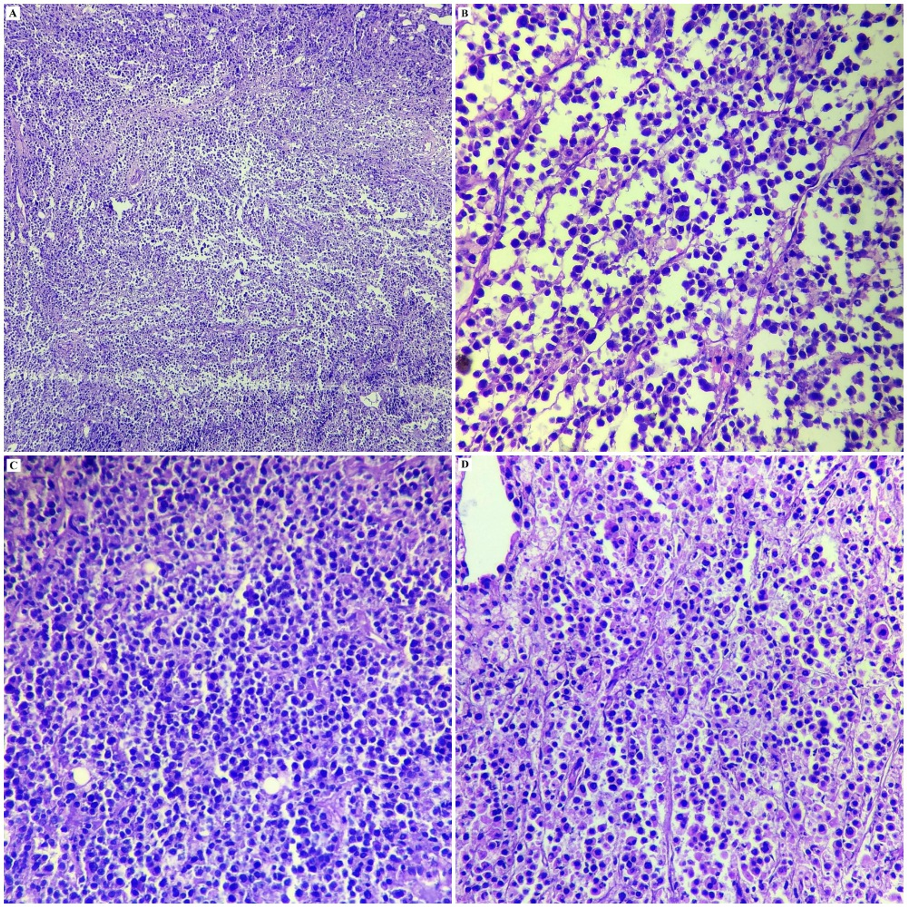
Frontiers | The Role of Histopathology as a Complementary ...
Detection of BrdU-label Retaining Cells in the Lacrimal Gland ... The photomicrographs depicted in Figure 2a-d show that following the 2- and 4-week chase periods, multiple BrdU + cells could be observed in any given lacrimal gland lobule. Figure 2a-g show that the number of BrdU + cells was high after 2 weeks of chase, decreased slightly by the 4 th week and then declined dramatically at 12 weeks.
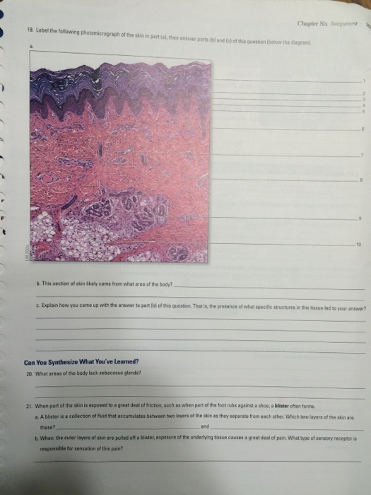
Solved Chapter Six Iniegumen 19 Label the following | Chegg.com
SC 2115 Anatomy and Physiology I - Mass These vary in name depending upon the tissue. 1. Fibroblasts are star-shaped cells responsible for the production of fibers and the matrix. 2. Fibrocytes are mature fibroblasts which maintain connective tissues. 3. Mast cells produce the anticoagulant, heparin, as well as th e irritating vasodilator, histamine. 4.
Cureus | Anaplastic Lymphoma Kinase Positive Large B-Cell ...
PDF Paper 1 Exam Questions (Live) - Mindset Learn The following table shows an analysis of the nutrients found in 100g portion of breakfast. The breakfast was ... Give two reasons by referring to the photomicrographs. (2) 2.2.2. Label structure 1. (1) 2.2.3. ... The photographs below show various human tissues 3.5. Identify each tissue type labeled A, ...

A photomicrograph of brown fat which is organized into ...

Single oral immunization of an attenuated Salmonella ...
Human induced pluripotent stem cells liver disease & toxicology

BSC2085L Lab 4 Exercises 5 & 6 Flashcards | Quizlet

Chapter 4: Tissues Flashcards | Quizlet
Get Answer) - Label the following photomicrographs with the ...

Exam 3 - Lab 5 Flashcards | Quizlet
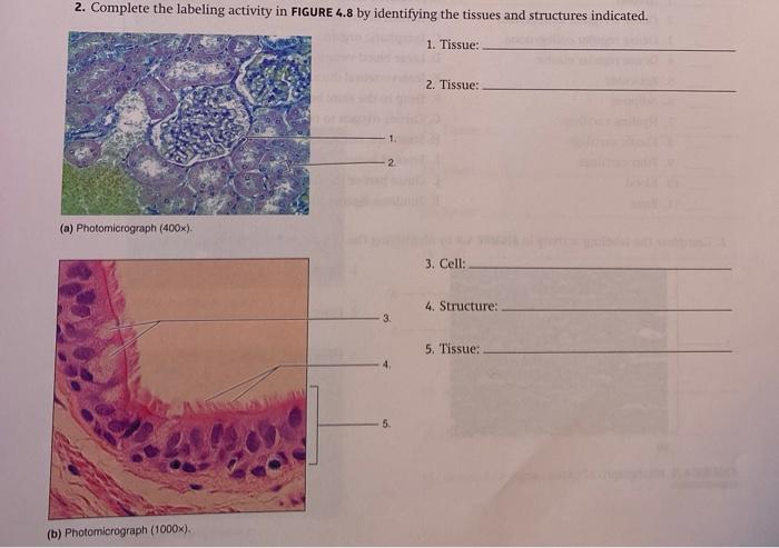
Solved 2. Complete the labeling activity in FIGURE 4.8 by ...
Lactobacillus casei HY2782 and Pueraria lobata Root Extract

Photomicrographs of Sertoli-NT2 cell tissue constructs formed ...
Photomicrographs High Resolution Stock Photography and Images ...

Myeloid caspase-8 restricts RIPK3-dependent proinflammatory ...

Chapter 4: Tissues Flashcards | Quizlet

Photomicrographs showing the variation in the amount of the ...
Portofolio Foto dan Gambar Stok dari Jubal Harshaw | Shutterstock

Photomicrographs and morphometric analysis of placental ...

NURR1 expression regulates retinal pigment epithelial ...

A bipotential organoid model of respiratory epithelium ...
Solved Experiment 2 Data Table 2 Photo 8 Et Photo 9 Photo 10 ...

Chapter 4: Tissues Flashcards | Quizlet
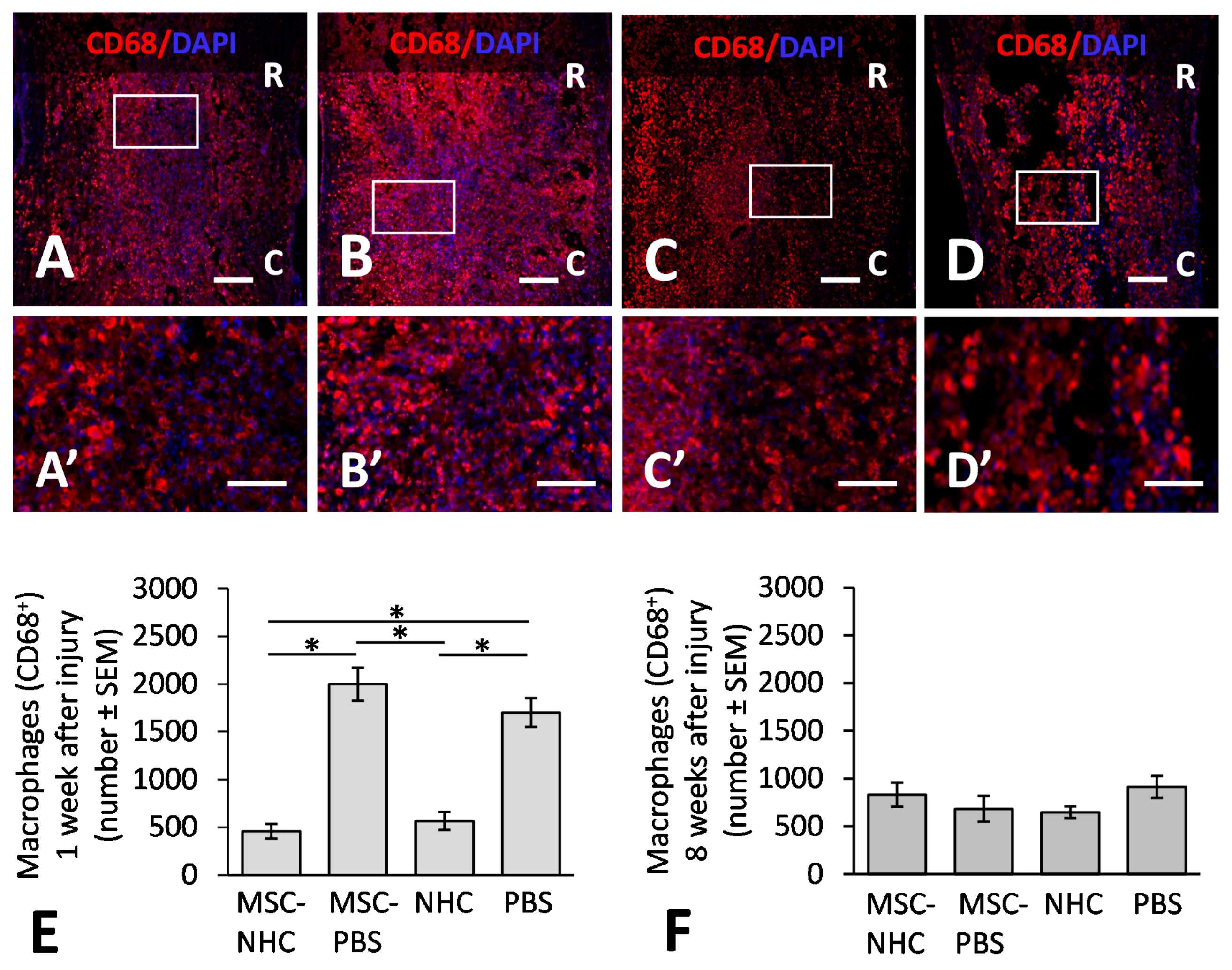
A) Representative photomicrographs of IBA1 (purple) and CD68 ...

Solved 25. Label the following photomicrographs by tissue ...

Tissues Chapter 5 Flashcards | Chegg.com

Solved EXERCISES Ties The muscles of your armare primarily ...

Chapter 4: Tissues Flashcards | Quizlet
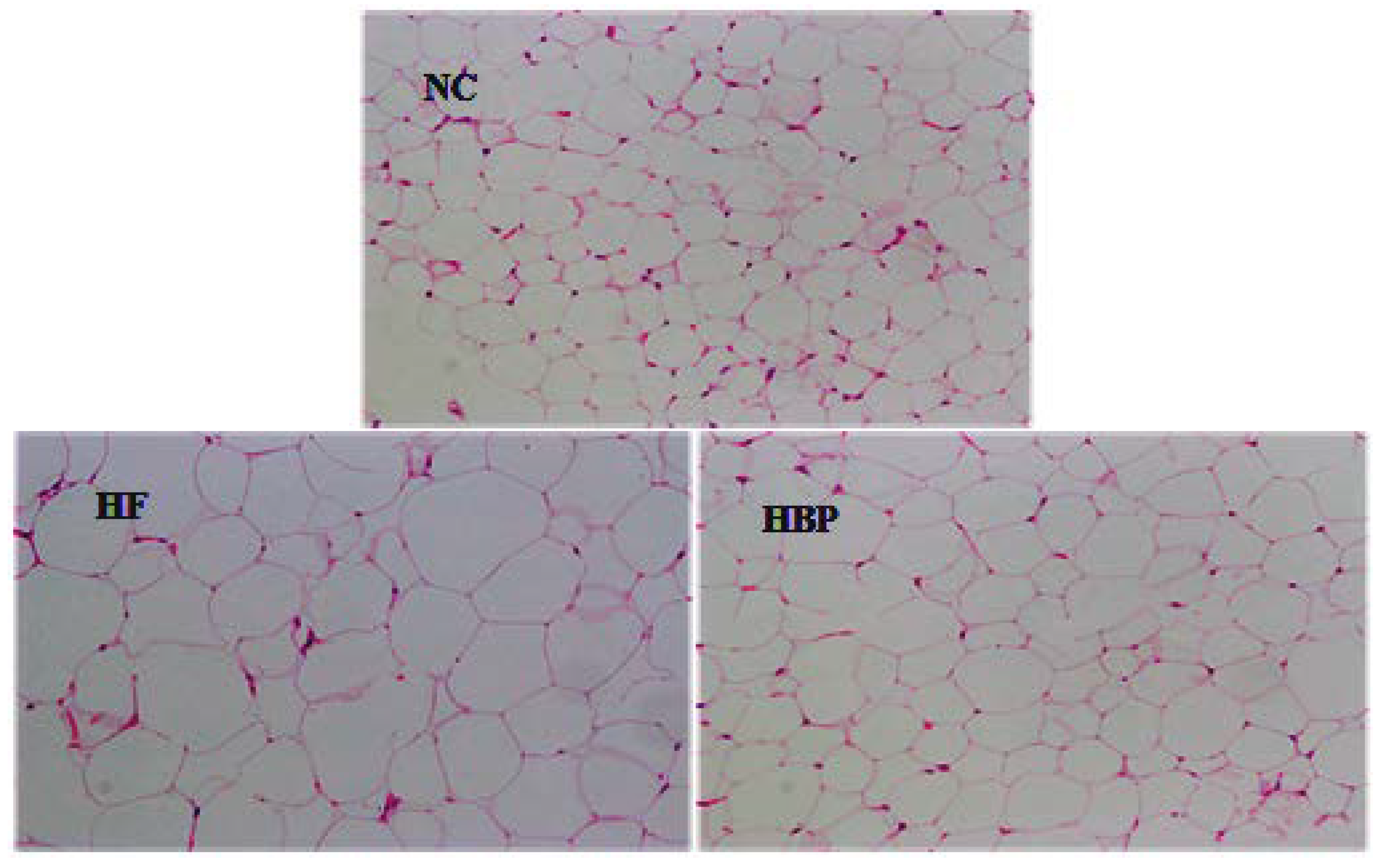
Nutrients | Free Full-Text | Hydrolyzed Bound Phenolics from ...
Cells | Free Full-Text | The Effects of the Combination of ...

Exam 3 - Lab 5 Flashcards | Quizlet

SOLUTION: Labact5 - Studypool

Liver - Cellink CN

Chapter 4: Tissues Flashcards | Quizlet
Post a Comment for "41 label the following photomicrographs by tissue type"