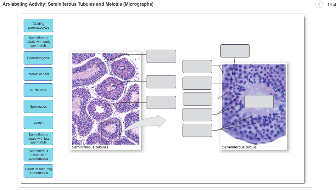
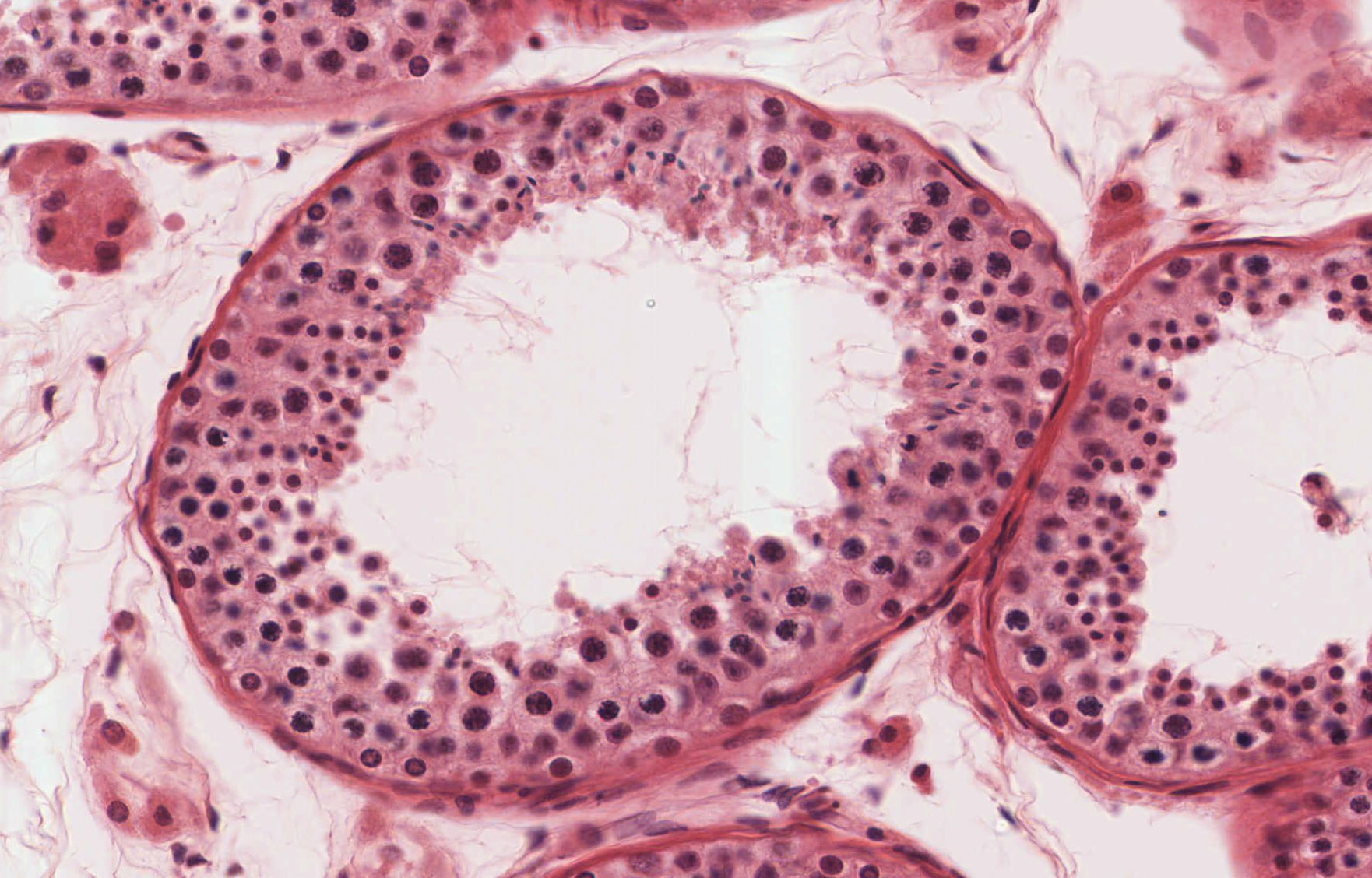
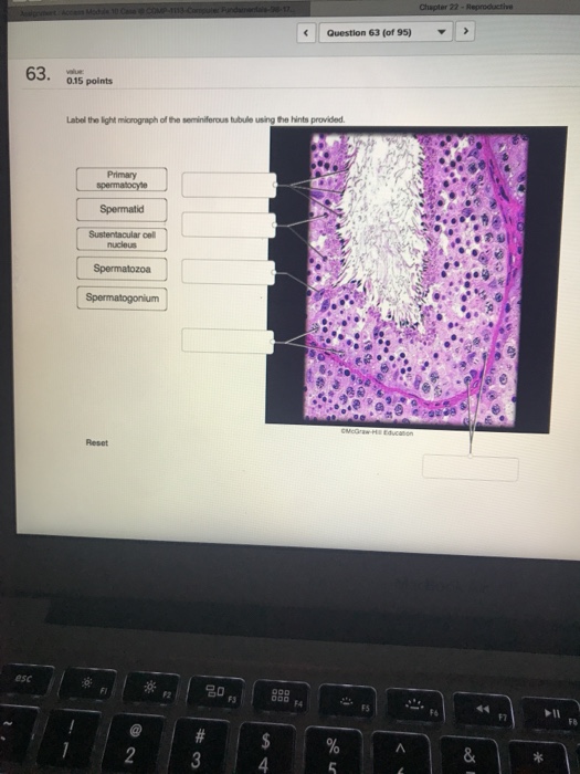
45 label the light micrograph of the seminiferous tubule using the hints provided
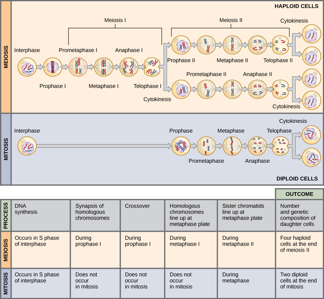
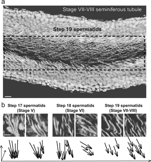
Abstracts from the 54 th European Society of Human Genetics (ESHG ... Enter the email address you signed up with and we'll email you a reset link. Epididymis Ductus deferens Ejaculatory duct Urethra Sperm (spermatozoa) production in seminiferous tubules ... Crossover is followed through the diagrams below. ... Scanning electron micrograph of a.
The Reproductive System - OERTX To fertilize an egg, sperm must be moved from the seminiferous tubules in the testes, through the epididymis, and—later during ejaculation—along the length of ...

Label the light micrograph of the seminiferous tubule using the hints provided
Label the light micrograph of the seminiferous tubule using the hints ... Label the light micrograph of the seminiferous tubule using the hints provided STUDY Learn Write Test PLAY Match Created by Sarah_Branning Terms in this set (4) lumen ... spermatid ... germ cell ... seminferous tubuke ... Male Reproductive System - Histology at the University of Michigan Returning to the slides of the seminiferous tubules, study the passageway by which the sperm pass from the seminiferous tubules through the rete testis, ... Reproductive lab Flashcards | Quizlet Ductus (vas) deferens Label the wall of the uterus using the hints provided. lumen of seminiferous tubules Epididymis Place the following structures of the male reproductive tract in order of how sperm passes through them to the external environment. endometrium ciliated simple columnar epithelium of uterine tube
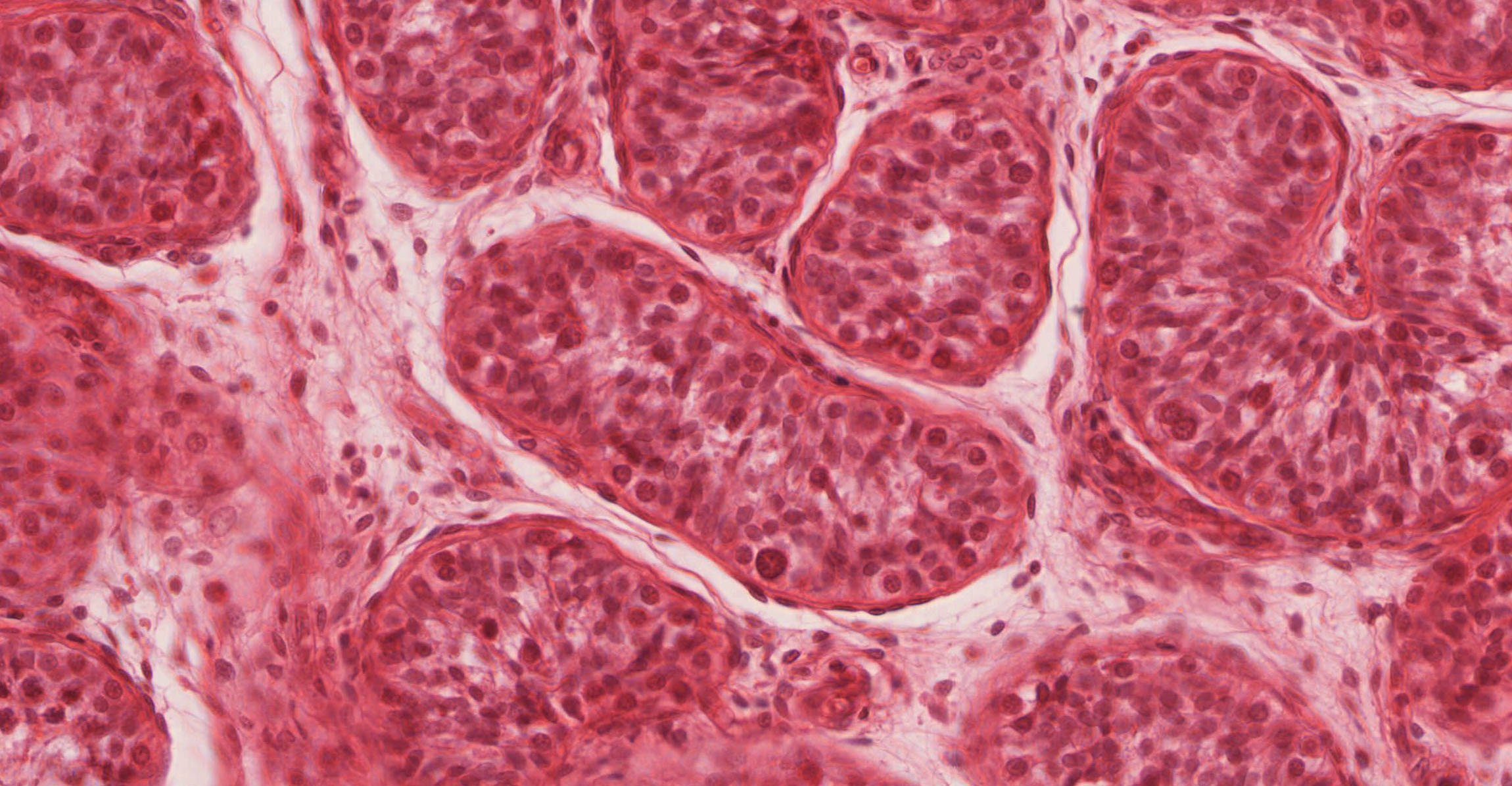
Label the light micrograph of the seminiferous tubule using the hints provided. Light Micrograph of a Seminiferous Tubule In Transverse Section Light Micrograph of a Seminiferous Tubule In Transverse Section. Variant Image ID: 14649. Add to Lightbox. Save to Lightbox. Email this page. Link this page. Print. Please describe! how you will use this image and then you will be able to add this image to your shopping basket. Anatomy and Physiology of the Male Reproductive System - BIO 140 ... Apr 18, 2022 ... (b) In this electron micrograph of a cross-section of a seminiferous tubule from a rat, the lumen is the light-shaded area in the center of ... Lab 10 Exercise 42 : Male reproductive Flashcards | Quizlet The ductus deferents is located within the Speematic cord Label the non-reproductive structures of the male pelvis using the hints provided Identify the parts of the penis in a cross section by clicking and dragging the labels to the correct location on the micrograph The seminiferous tubules produce Sperm Electron micrograph of seminiferous tubules in October. Sertoli cell... This present study investigated the seasonal morphological changes in SCs in the reproductive cycle of Pelodiscus sinensis by light microscopy, transmission ...
Ch 27 Lab Flashcards | Quizlet Label the testis and spermatic cord using the hints provided. 1. The ovaries are paired, oval-shaped organs found in the pelvic cavity. 2. The uterine tubes receive the ovulated oocyte and are the most common place for fertilization to take place. 3. The uterus is a pear shaped muscular organ where fetal development takes place. 4. Solved Label the testis and spermatic cord using the hints - Chegg Expert Answer. 100% (1 rating) Ans: Left side: top to bottom 1) vas deferenc …. View the full answer. Transcribed image text: Label the testis and spermatic cord using the hints provided. Testis Visceral layer of tunica vaginalis Pampiniform plexus Head Testicular artery Tail Vas deferens Epididymis Body Reset Zoom. Reproductive System 6. Detail the gross and microscopic anatomy of the mammary glands. A sagittal section through the female pelvis illustrates the internal reproductive structures ... Light Micrograph of the Wall of a Seminiferous Tubule Images on Similar Topics · Anatomy · Histology · Light · Male Reproductive System · Microanatomy · Micrograph · Seminiferous · Seminiferous tubules
Ch 27 Reproductive Flashcards | Quizlet Cells arrested in metaphase, when chromosomes are most highly condensed, are stained and then viewed with a microscope equipped with a camera. A photo is displayed and the images of the chromosomes are arranged into pairs according to their appearance. • This karyotype shows the chromosomes from a normal human male. Homologous Chromosomes/Homologs EOF Solved Label the light micrograph of the semintferous tubule - Chegg Expert Answer 86% (7 ratings) A … View the full answer Transcribed image text: Label the light micrograph of the semintferous tubule using the hints provided. Sustentacular cell Primary spermatocyte Spermatozoon Lumen Fibromuscular wall Spermatogonium Basal lamina Spermatid Seminferous tubule BTC Reglare Co r e Reset Zoom Reproductive lab Flashcards | Quizlet Ductus (vas) deferens Label the wall of the uterus using the hints provided. lumen of seminiferous tubules Epididymis Place the following structures of the male reproductive tract in order of how sperm passes through them to the external environment. endometrium ciliated simple columnar epithelium of uterine tube
Male Reproductive System - Histology at the University of Michigan Returning to the slides of the seminiferous tubules, study the passageway by which the sperm pass from the seminiferous tubules through the rete testis, ...
Label the light micrograph of the seminiferous tubule using the hints ... Label the light micrograph of the seminiferous tubule using the hints provided STUDY Learn Write Test PLAY Match Created by Sarah_Branning Terms in this set (4) lumen ... spermatid ... germ cell ... seminferous tubuke ...











Post a Comment for "45 label the light micrograph of the seminiferous tubule using the hints provided"