44 labeling microscope parts
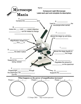
Microscope Parts and Lenses Trivia Quiz | Miscellaneous Science | 10 ... The lower the magnitude of the microscope lenses, the lower the light needs to be, and the higher the magnitude, the higher the light. 9. If you have one lens of your microscope with 4X, and the other with 10X, what is the total multiplication? Answer: 40. The rule of microscope magnification is simple. Microscope Parts & Specifications | Microscope World Resources If your microscope has a fine focus adjustment, turning it a bit should be all that's necessary. Continue with subsequent objective lenses and fine focus each time. If you are unsure of the parts and functions of your microscope, contact Microscope World. This page has activities and free printouts for labeling parts of the microscope.
Microscope: Types of Microscope, Parts, Uses, Diagram - Embibe Any microscope consists of three parts: Head (contains the optical parts), base( supports the structure) and arm(connects the head to the base). The eyepiece lens is closer to the observer's eye. In comparison, the objective lens, which gives a \(400 - 100\) times magnified image, is closer to the object.

Labeling microscope parts
› products › microscopeMicroscope Parts & Accessories | Products | Leica Microsystems Sep 23, 2019 · Microscope Parts & Accessories. Leica Microsystems provides a broad range of microscope parts and accessories to perfectly tailor your imaging system for your needs and budget. View our catalog of illumination, objectives, filter cubes, filter wheels and more. Metaphase - Genome.gov Metaphase is a stage during the process of cell division (mitosis or meiosis). Normally, individual chromosomes are spread out in the cell nucleus. During metaphase, the nucleus dissolves and the cell's chromosomes condense and move together, aligning in the center of the dividing cell. At this stage, the chromosomes are distinguishable when ... Search | Leica Microsystems 4 October 2022, Wetzlar, Germany - Leica Microsystems, a leader in microscopy and scientific instrumentation, has launched Enersight, an intuitive new software platform for microscope inspection and quality control. The software is designed to simplify and streamline the entire inspection and docume. News - 29 Mar 2022.
Labeling microscope parts. Cleanliness Analysis with a 2-methods-in-1 solution In this article, it is examined how an overall efficient and cost-effective cleanliness analysis workflow can be achieved with a 2-methods-in-1 materials analysis solution, combining optical microscopy and laser induced breakdown spectroscopy (LIBS).Technical cleanliness is important for assuring product quality and reliability in the automotive and electronics industries. leaf | Definition, Parts, & Function | Britannica The main function of a leaf is to produce food for the plant by photosynthesis. Chlorophyll, the substance that gives plants their characteristic green colour, absorbs light energy. The internal structure of the leaf is protected by the leaf epidermis, which is continuous with the stem epidermis. The central leaf, or mesophyll, consists of soft ... Microscope slide - Wikipedia A microscope slide is a thin flat piece of glass, typically 75 by 26 mm (3 by 1 inches) and about 1 mm thick, used to hold objects for examination under a microscope.Typically the object is mounted (secured) on the slide, and then both are inserted together in the microscope for viewing. This arrangement allows several slide-mounted objects to be quickly inserted and … Microscope Labeling Game - PurposeGames.com This is an online quiz called Microscope Labeling Game. There is a printable worksheet available for download here so you can take the quiz with pen and paper. This quiz has tags. Click on the tags below to find other quizzes on the same subject. Science. microsope. Your Skills & Rank. Total Points. 0. Get started! Today's Rank--0. Today 's Points. One of us! Game Points. …
5 White Blood Cells Types and Their Functions - New Health Advisor Agranulocytes are free of visible grains under the microscope and include lymphocytes and monocytes. Together, they coordinate with one another to fight off things like cancer, cellular damage, and infectious diseases. Below, detailed information about each type will be discussed. 1. Neutrophils Service & Support | Leica Microsystems Our commitment to you as a Leica customer is to deliver best-in-class services entirely dedicated to microscopy. With over 170 years of history, nobody knows your microscope better than Leica. We provide complete workflow support to enable your success through unmatched expertise. Spinal Cord Cross Section | New Health Advisor Basically the spinal cord is responsible for: The electrical communication between the various parts of the body and the brain. The spinal cord runs through several different levels of the trunkmaking it easier to communicate as the electrical signals are conducted through the cord. Walking, also referred to as locomotion. Medicine & Health | UNSW Sydney Research & impact. UNSW Medicine & Health is world renowned for its research and impact addressing issues of health disparities and improving lives through our key research themes of Cancer; Infectious Disease, Immunity & Inflammation; Neuroscience, Mental Health & Addiction; Cardiac, Vascular & Metabolic Medicine and Health Systems.
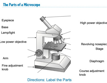
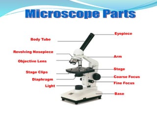
› t-partsMicroscope Parts & Specifications | Microscope World Resources If your microscope has a fine focus adjustment, turning it a bit should be all that's necessary. Continue with subsequent objective lenses and fine focus each time. If you are unsure of the parts and functions of your microscope, contact Microscope World. This page has activities and free printouts for labeling parts of the microscope. laboratoryinfo.com › labeling-parts-of-the-microscopeParts of the Microscope with Labeling (also Free Printouts) Mar 07, 2022 · Click to Download : Label the Parts of the Microscope with answers (A4) PDF print version.. For a thorough review of each microscope part continue reading….. A basic microscope has a single convex lens such as those found in a magnifying glass, which you can use to visualize the finest prints. Parts of a Microscope with Their Functions - Microbe Online The arm as well as the base help to carry the microscope. In the case of high-quality microscopes, an articulated arm with more than one joint is present. Base. The base is the bottom of a microscope. It helps to support the microscope. A microscopic illuminator is also present in it. Microscope Worksheet My staining didn't work part 1: Background staining in IHC and IF A particular nuisance in immunostaining is the appearance of background staining, which is the non-specific staining of tissue and cell elements by colorimetric or fluorescence dyes. Researchers have difficulty identifying the source of the background staining because IHC and IF experiments involve multiple reagents including the tissue ...
Recombinant Anti-p27 KIP 1 antibody [Y236] (ab32034) Immunocytochemistry/ Immunofluorescence analysis of RAW264.7(Mouse Abelson murine leukemia virus-induced tumor macrophage) treated with 100ng/ml LPS for 7 h and 1µg/ml BFA for the last 3h cells labeling p27 KIP 1 with purified ab32034 at 1/100 dilution(5.22 µg/ml). Cells were fixed in 4% paraformaldehyde and permeabilized with 0.1% Triton X-100.
Autoclave: Principle, Procedure, Types, Uses - Microbe Online Types of Autoclaves. There are different types of autoclaves available. Gravity displacement type autoclave: It is the most common type used in laboratories and is available in various sizes and dimensions. Vertical type (small volume capacity) Horizontal autoclave (large volume capacity) Positive pressure displacement type autoclave.
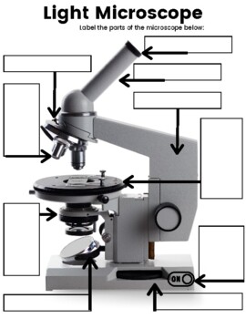
Microscope Labeling - The Biology Corner Students label the parts of the microscope in this photo of a basic laboratory light microscope. Can be used for practice or as a quiz. Name_____ Microscope Labeling . Microscope Use: 15. When focusing a specimen, you should always start with the _____ objective. 16. When using the high power objective, only the _____ knob should be used. 17. The type of microscope used …
› game › microscope-labelingMicroscope Labeling Game - PurposeGames.com This is an online quiz called Microscope Labeling Game. There is a printable worksheet available for download here so you can take the quiz with pen and paper. This quiz has tags. Click on the tags below to find other quizzes on the same subject.
Microscope Products | Leica Microsystems Microscope Products We develop high-tech precision microscopes for the analysis of microstructures with the user, for the user. In our product portfolio you will find microscopes for life science including biotechnology and medicine as well as for research and development of raw materials and industrial quality assurance.
Cell Division: Mitosis and Meiosis - Owlcation Cell division is the process by which biological cells multiply. There are three major types of cell division: Mitosis - used by Eukaryotic organisms to grow or reproduce asexually;; Meiosis - used by Eukaryotic organisms to create sex cells (gametes);; Binary Fission - used by Prokaryotic organisms to reproduce.; Despite their differences, remember that all three types of cell division begin ...
Mr. Jones's Science Class Parts of the Atom Creating Atomic Models Isotopes and Ions Practice Scanning Tunneling Microscope (STM) Find Someone Who... - Atoms Scientists Structure of the Atom - Research Scientist Presentation (PPT.) Presentation Rubric - Scientist Point of View Guide History of the Atom - Scientists - Resource Sheet History of the Atom (PPT.)
Experiments Show How Difficult It Is to Infect Others with Influenza ... Professor Carl Heneghan and Tom Jefferson highlighted how difficult it is for someone to transmit an influenza-like illness ("ILI") to another person. They highlight experiments, or challenge studies, done long before Covid with rhinoviridae which causes common colds. "If you have followed these experiments and paid attention, just like the authors, you might conclude how…
Home Lab Beginners guide - Hardware - Hayden James A rack unit (abbreviated U or RU) is a unit of measure defined as 1 3⁄4 inches (or 44.45 mm). It's the unit of measurement for the height of 19-inch and 23-inch rack frames and the equipment's height. The height of the frame/equipment is expressed as multiples of rack units. A typical full-size rack is 42U high.
Light Microscope (Theory) - Amrita Vishwa Vidyapeetham Parts of a Microscope . It consists of mainly three parts: Mechanical part - base, c-shaped arm and stage. Magnifying part - objective lens and ocular lens. Illuminating part - sub stage condenser, iris diaphragm, light source. Mechanical part. Base: It helps in holding the various parts of microscope. It also contains the light source.
Golgi Body - Genome.gov Definition. 00:00. …. A Golgi body, also known as a Golgi apparatus, is a cell organelle that helps process and package proteins and lipid molecules, especially proteins destined to be exported from the cell. Named after its discoverer, Camillo Golgi, the Golgi body appears as a series of stacked membranes.
Recombinant Anti-CD161 antibody [EPR26340-6] (ab302564) Image acquisition was performed with Leica SP8 confocal microscope. Multiplex immunohistochemistry - Anti-CD161 antibody [EPR26340-6] (AB302564) Multiplex immunohistochemical analysis of formalin/PFA-fixed paraffin-embedded sections of human tonsil tissue labeling CD-161 with AB302564 at 1:100 dilution (4.96 μg/mL) colored in red (Opal™570 ...
Gram Stain Technique - Amrita Vishwa Vidyapeetham Drawing a circle on the underside of the slide using a glassware-marking pen may be helpful to clearly designate the area in which you will prepare the smear. You may also label the slide with the initials of the name of the organism on the edge of the slide. Care should be taken that the label should not be in contact with the staining reagents.
Diaphragm: Location, anatomy, innervation and function | Kenhub The diaphragm is an unpaired, dome shaped skeletal muscle that is located in the trunk. It separates the thoracic and abdominal cavities from each other by closing the inferior thoracic aperture. The diaphragm is the primary muscle that is active in inspiration. Contraction of the muscle facilitates expansion of the thoracic cavity.
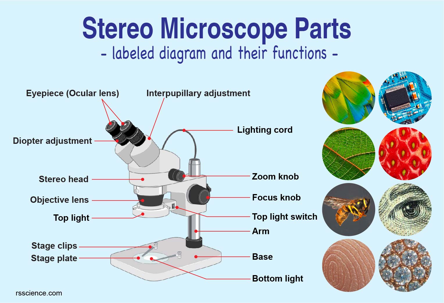
Binocular Microscope Anatomy - Parts and Functions with a Labeled ... The 12 parts of the microscope include - illuminators, condenser, iris diaphragm, objective lens, eyepieces, body tubes, arm or frame, coarse adjustment, fine adjustments, nose piece, stage, and base.
› game › 5f76372de3Microscope Parts Quiz - PurposeGames.com Nov 04, 2009 · This is an online quiz called Microscope Parts There is a printable worksheet available for download here so you can take the quiz with pen and paper. Your Skills & Rank
scheme work biology - Free KCPE Past Papers Draw and label the light microscope; Description of a cell; Drawing and labeling the light microscope . Light microscope; ... Golden tips Biology Page 15-16; KLB teachers book 1 pages 23-25 . 10. 1-2. THE CELL. Parts of the light microscope and their functions . Calculation of magnification using light microscope. By the end of the lesson, the ...
KEYENCE TV : XM-5000 - Handheld CMM | KEYENCE America XM-5000 - Handheld CMM 2:20. 3D Surface Profiler | VK-X3000 Series 2:10. All-in-One Fluorescence Microscope | BZ-X Series 2:22. Telecentric Measurement System - TM-X5000 Series 2:13. Accurate and Traceable 2D Measurement with TM-X5000 Series 1:39. Flexible, Calibrated Alternative to Backlit Measurement with TM-X5000 Series 1:21.
13 Best Image Annotation Tools of 2022 [Reviewed] - V7Labs Hive Data is a fully managed data annotation solution to source and label training data for AI / ML Models. Hive Data supports image, video, text, 3D Point Cloud annotation and data sourcing. Apart from basic annotation types, Hive Data offers multi-frame object tracking, contours, and 3D panoptic segmentation.
› microscope_labelingMicroscope Labeling - The Biology Corner 18. You should carry the microscope by the _____ and the _____. 19. The objectives are attached to what part of the microscope (it can be rotated to click lenses into place?) _____ 20. A microscope has an ocular objective of 10x and a high power objective of 50x, what is the microscope's total magnification?
TMZ Sports | Latest News, Videos & Photos TMZ Sports posted video Friday showing the NBA star punching Poole in the face at practice Wednesday in San Francisco, after the two men got into a verbal argument. Draymond says he watched the ...
Carl Zeiss Meditec, Inc. - Medical Solutions made by ZEISS - Medical ... Not all products, services or offers are approved or offered in every market and approved labeling and instructions may vary from one country to another. For country specific product information, see the appropriate country website. Product specifications are subject to change in design and scope of delivery as a result of ongoing technical ...
› t-labeling_microscopeLabeling the Parts of the Microscope | Microscope World Resources Labeling the Parts of the Microscope. This activity has been designed for use in homes and schools. Each microscope layout (both blank and the version with answers) are available as PDF downloads.
Tinea Manuum: Causes, Symptoms, and Treatment Tinea Manuum Symptoms. Tinea manuum is an itchy hand rash that is caused by a fungal infection. The symptoms include: 1. Itchy fingers and hands. Redness. Flaky or peeling skin. Ring-shaped rash. The infection can occur on one or both hands but usually only develops on one hand.
Search | Leica Microsystems 4 October 2022, Wetzlar, Germany - Leica Microsystems, a leader in microscopy and scientific instrumentation, has launched Enersight, an intuitive new software platform for microscope inspection and quality control. The software is designed to simplify and streamline the entire inspection and docume. News - 29 Mar 2022.
Metaphase - Genome.gov Metaphase is a stage during the process of cell division (mitosis or meiosis). Normally, individual chromosomes are spread out in the cell nucleus. During metaphase, the nucleus dissolves and the cell's chromosomes condense and move together, aligning in the center of the dividing cell. At this stage, the chromosomes are distinguishable when ...
› products › microscopeMicroscope Parts & Accessories | Products | Leica Microsystems Sep 23, 2019 · Microscope Parts & Accessories. Leica Microsystems provides a broad range of microscope parts and accessories to perfectly tailor your imaging system for your needs and budget. View our catalog of illumination, objectives, filter cubes, filter wheels and more.












Post a Comment for "44 labeling microscope parts"