41 provide the labels for the electron micrograph
The Apex of Protein Tags for Electron Microscopy The Apex of Protein Tags for Electron Microscopy October 22, 2012 0 Scientists have developed a new peroxidise-derived tag that can be used to label proteins for visualization by electron... Plant Cell Nucleus Electron Micrograph : Cell And Organelles Dr Jastrow ... Below is a collection of electron micrographs with labelled subcellular structures that you should be able to identify. In mammals it's average diameter is about 6 an electron micrograph of a section through an animal cell nucleus (from an insect cell). In flowering plants, this condition occurs in sieve tube elements.74.
Solved Label the transmission electron micrograph based on - Chegg Solved Label the transmission electron micrograph based on | Chegg.com. Science. Biology. Biology questions and answers. Label the transmission electron micrograph based on the hints provided Mitochondrion Heterochromatin Plasma cell Nucleus Rough endoplasmic reticulum Nucleolus.

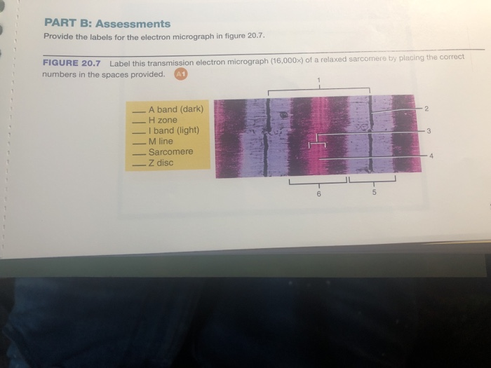
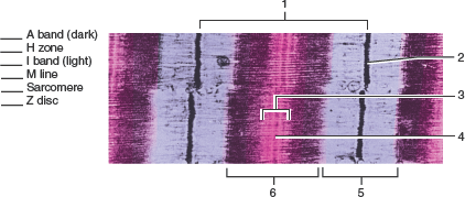
Provide the labels for the electron micrograph
Electron Micrographs of Cell Organelles | Zoology - Biology Discussion This is an electron-micrograph of plastid or chloroplast, which is an integral component of all green plant leaves and is characterized by following features (Fig. 15 & 16): (1) They may be spheroidal, ovoid, stellate or collar shaped and differ in size and number in different cells. What is Electron Microscopy? - UMASS Medical School Electron microscopy (EM) is a technique for obtaining high resolution images of biological and non-biological specimens. It is used in biomedical research to investigate the detailed structure of tissues, cells, organelles and macromolecular complexes. The high resolution of EM images results from the use of electrons (which have very short ... chloroplast and mitochondrion.pdf - D SBA#: Title: Electron micrograph ... At least 2 parts labeled correctly (1 mark) 4 Annotate drawings appropriately and accurately e. 5 parts annotated correctly (4 marks)f. 4 parts annotated correctly (3 marks) g. 3 parts annotated correctly (2 marks)h.
Provide the labels for the electron micrograph. What Is an Electron Microscope (EM) and How Does It Work? - VHA ... A stream of high voltage electrons (usually 5-100 KeV) is formed by the Electron Source (usually a heated tungsten or field emission filament) and accelerated in a vacuum toward the specimen using a positive electrical potential. This stream is confined and focused using metal apertures and magnetic lenses into a thin, focused, monochromatic beam. Electron Micrographs - University of Oklahoma Health Sciences Center Below is a collection of electron micrographs with labelled subcellular structures that you should be able to identify. Also, be sure to observe any electron micrographs which are made available in the laboratory by the instructor. Electron microscope - Wikipedia An electron microscope is a microscope that uses a beam of accelerated electrons as a source of illumination. As the wavelength of an electron can be up to 100,000 times shorter than that of visible light photons, electron microscopes have a higher resolving power than light microscopes and can reveal the structure of smaller objects.. Electron microscopes use shaped magnetic … Light and Electron Microscopy Flashcards | Quizlet A microscope that allows light rays to pass directly to the eye without being deflected by an intervening opaque plate in the condenser. Light Source Provides illumination of variable intensity Condenser Lens Focuses a cone of light on the specimen Condenser Diaphragm Controls the size of the cone of light that reaches the specimen Stage
Label This Transmission Electron Micrograph Of A Relaxed ... - Blogger Provide the labels for the electron micrograph in figure 18.5. (b) section through a muscle in the extended condition (140 % of whole muscle resting length). Label the following image using the terms provided. Label this transmission electron micrograph of relaxed sarcomeres by clicking and dragging the labels to the correct location . Ultrasensitive detection of patulin based on a Ag+-driven one ... After the formation of stable Au-N bonds between AuNFs and g-C 3 N 4 nanosheets by chemical bonding, the typical transmission electron micrograph of the AuNFs/g-C 3 N 4 composite in Fig. 1 G shows that flower-like particles are fixed on a single nanosheets, indicating that AuNFs were successfully loaded onto the surface of g-C 3 N 4 nanosheets. Looking at the Structure of Cells in the Microscope ... Moreover, in recent years, the performance of electron microscopes has been improved by the development of electron illumination sources called field emission guns. These very bright and coherent sources can substantially improve the resolution achieved. The major landmarks in the development of electron microscopy are listed in Table 9-2. Label the microscope — Science Learning Hub Use this with the Microscope parts activity to help students identify and label the main parts of a microscope and then describe their functions. Drag and drop the text labels onto the microscope diagram. If you want to redo an answer, click on the box and the answer will go back to the top so you can move it to another box.
What is Electron Microscopy? - UMASS Medical School It is termed a scanning electron microscope because the image is formed by scanning a focused electron beam onto the surface of the specimen in a raster pattern. The interaction of the primary electron beam with the atoms near the surface causes the emission of particles at each point in the raster (e.g., low energy secondary electrons, high energy back scatter electrons, X-rays … Solved Please label the electron micrograph to assess your | Chegg.com Question: Please label the electron micrograph to assess your knowledge of the structure and function of a cell's nucleus nuclear pore endoplasma reticulum chromatin nucleolus nuclear envelope . This problem has been solved! See the answer See the answer See the answer done loading. Chapter 2 Correlated light and electron microscopy/electron tomography ... Abstract. Three-dimensional light microscopy and three-dimensional electron microscopy (electron tomography) separately provide very powerful tools to study cellular structure and physiology, including the structure and physiology of mitochondria. Fluorescence microscopy allows one to study processes in live cells with specific labels and ... Electron Microscopy Sciences Expiration Labels - Fisher Sci Manufacturer: Electron Microscopy Sciences 7702405 Quickly identify outdated laboratory items such as reagents and controls. These labels provide space for noting the date that the items were received, opened and when it will expire. A permanent adhesive holds the label firmly in place on a variety of surfaces. The label measures 3/4" x 1 1/2".
Muscle Lab 19 Figure 19.5 Sarcomere Diagram | Quizlet
Electron Microscope — Penn State These labels are generated based on the underlying awards/grants. Together they form a unique fingerprint. electron microscopesAgriculture & Biology100% microscopesAgriculture & Biology78% development aidAgriculture & Biology56% digital imagesAgriculture & Biology47% instrumentationAgriculture & Biology46%

Scanning electron micrograph of the labella of horn fly ...
Identify The Bands And Lines Of The Striations Of A Tem Of ... 15 Dec 2020 — Provide the labels for the electron micrograph in figure 18.5. Figure 18.5 Identify the bands and lines of the striations in this ...

Electron micrograph of an asymmetric synapse from dissociated ...
Renal System | histology - University of Michigan Correct answer 4. A podocyte - Note that you can see areas where the cytoplasm of the cell is connected to the pedicels (foot processes, labeled "p"). "V" labels vascular lumen. "E" indicates endothelial cell cytoplasm. "f" indicates an endothelial fenestra. The bracket indicates basal lamina. "U" indicates urinary space.

Solved Mitochondrion Nucleus Vesicle Peroxisome | Chegg.com
Renal System | histology - University of Michigan Note that we do not expect you to be able to distinguish among these 3 cell types by light microscopy. The parietal layer of Bowman's capsule is also a simple squamous epithelium which transitions to cuboidal epithelium of the proximal convoluted tubule at the urinary pole slide 210 View Image.Look around under low power to find glomeruli sectioned through the vascular pole.
animal cell electron micrograph labelling Diagram | Quizlet
Optical Metasurfaces for Energy Conversion | Chemical Reviews Nanostructured surfaces with designed optical functionalities, such as metasurfaces, allow efficient harvesting of light at the nanoscale, enhancing light–matter interactions for a wide variety of material combinations. Exploiting light-driven matter excitations in these artificial materials opens up a new dimension in the conversion and management of energy at the nanoscale. In …

Scanning electron micrographs illustrating lobed chlorenchyma ...
Multicolor Labels in Electron Microscopy [Roger Tsien ... - BioTechniques Lab design and machinery. After 15 years of research, the late, Nobel Prize-winning, scientist Roger Tsien and his colleagues bring multicolor labels to electron microscopy. Three new discoveries have already emerged. Every year over Christmas break, Roger Tsien gave himself a treat: he spent a few uninterrupted weeks in the lab working on ...
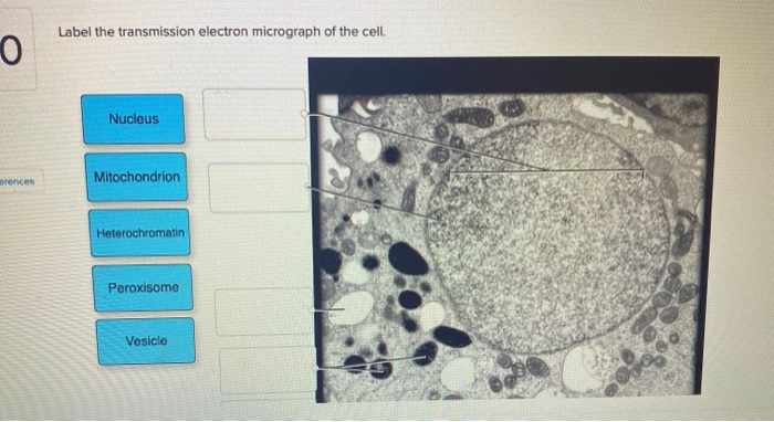
Solved Label the transmission electron micrograph of the ...
Color highlights detail in electron microscope images - New Atlas Color highlights detail in electron microscope images. By Michael Irving. November 08, 2016. On the left is a standard grayscale electron micrograph of a mouse brain, and on the right is a ...

Skeletal Muscle EM
Electron Microscope - an overview | ScienceDirect Topics A. Scholl, in Encyclopedia of Materials: Science and Technology, 2002 1 Photoemission Electron Microscopy. PEEM was first used in the 1930s and has since then matured into an established surface science technique (Stöhr et al. 1993, Tonner et al. 1995, Stöhr et al. 1998, Anders et al. 1999).PEEM is closely related to the Low Energy Electron Microscope (LEEM) and the Spin-polarized Low-energy ...

Pre-embedding immunogold labeling to optimize protein ...
Ultra-Bright and Stable Luminescent Labels for Correlative ... Correlative cathodoluminescence electron microscopy (CCLEM) bioimaging has recently been suggested to provide an attractive alternative based on labels emitting characteristic light. While luminescence excitation by an electron beam enables sub-diffraction imaging, structural damage to the sample by high energy electrons has been identified as ...

This scanning electron micrograph (SEM) depicted a number of ...
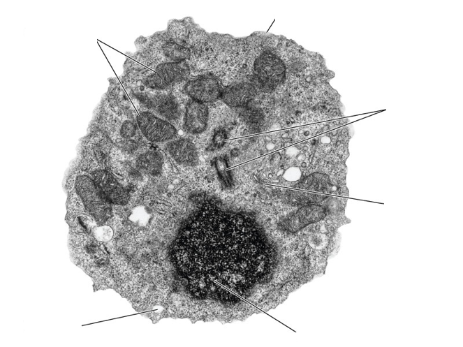
PDF Identifying Organelles from an Electron Micrograph - Ms JMO's Biology ... Courtesy of Dr. Julian Thorpe - EM & FACS Lab, Biological Sciences University Of Sussex The electron micrograph displayed below illustrates many of the plant cell characteristics discussed The cell wall, large central vacuole and chloroplasts are clearly visible Also visible is the clearly defined nucleus containing chromatin

BSC2085L Lab 8 Exercises 12, 13, & 14 Flashcards | Quizlet
The Mitochondrion - Molecular Biology of the Cell - NCBI … Mitochondria occupy a substantial portion of the cytoplasmic volume of eucaryotic cells, and they have been essential for the evolution of complex animals. Without mitochondria, present-day animal cells would be dependent on anaerobic glycolysis for all of their ATP. When glucose is converted to pyruvate by glycolysis, only a very small fraction of the total free energy …

Transmission electron micrographs showing phloem cells of ...
Electron microscope - Wikipedia An electron microscope is a microscope that uses a beam of accelerated electrons as a source of illumination. As the wavelength of an electron can be up to 100,000 times shorter than that of visible light photons, electron microscopes have a higher resolving power than light microscopes and can reveal the structure of smaller objects.

Immunogold-labeling of glutathione. Transmission electron ...
Oncology Reports - Spandidos Publications If labels cannot fit on the 17‐cm‐wide page unless the font size is smaller than 8 points, the figure must be split into several parts. Font style and appearance; Labels must be saved using standard fonts (Times New Roman, Times, Arial, Helvetica or Symbol font). The labels should be of the same font and size in all figures.

Transmission electron micrograph of an animal cell - Stock ...
The Mitochondrion - Molecular Biology of the Cell - NCBI ... The process of electron transport begins when the hydride ion is removed from NADH (to regenerate NAD +) and is converted into a proton and two electrons (H-→ H + + 2e-). The two electrons are passed to the first of the more than 15 different electron carriers in the respiratory chain. The electrons start with very high energy and gradually ...

Electron micrograph of a rabbit retina grown in situ (p"14 ...
Diagram of labeling used for light and electron microscopy. A: Labeling ... An ideal label for light and electron microscopy would contain both fluorescent dye and a colloidal nanoparticle on a single antibody molecule. Secondary and tertiary label, the colloidal ...
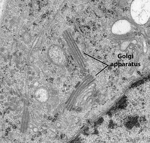
BIOL 230 Lecture Guide - Electron Micrograph of a Golgi Body
Electron Microscope- Definition, Principle, Types, Uses, Labeled Diagram Electron Microscope is in the form of a tall vacuum column that is vertically mounted. It has the following components: 1. Electron gun The electron gun is a heated tungsten filament, which generates electrons. 2. Electromagnetic lenses The condenser lens focuses the electron beam on the specimen.

Labeling the Cell Flashcards | Quizlet
Optical Sensor - an overview | ScienceDirect Topics For example, the linkage of an anthracene fluorophore with a crown ether receptor creates a diamine sensor for detecting food spoilage. 52 In the absence of guest molecules (diamines such as putrescine or cadaverine), the fluorescence of anthracene is “switched-off” by photo-induced electron transfer; when a diamine is bound, however ...

A representative electron micrograph illustrating the ...
Animal Cell Electron Microscope Labelled - Q14 Draw a large diagram of ... Here is an electron micrograph of an animal cell with the labels superimposed: Make your work easier by using a label. Make your work easier by using a label. After this, add another oval shape outside the line you just drew, and this will make the cell membrane to your animal cell. You see that many features are in common.
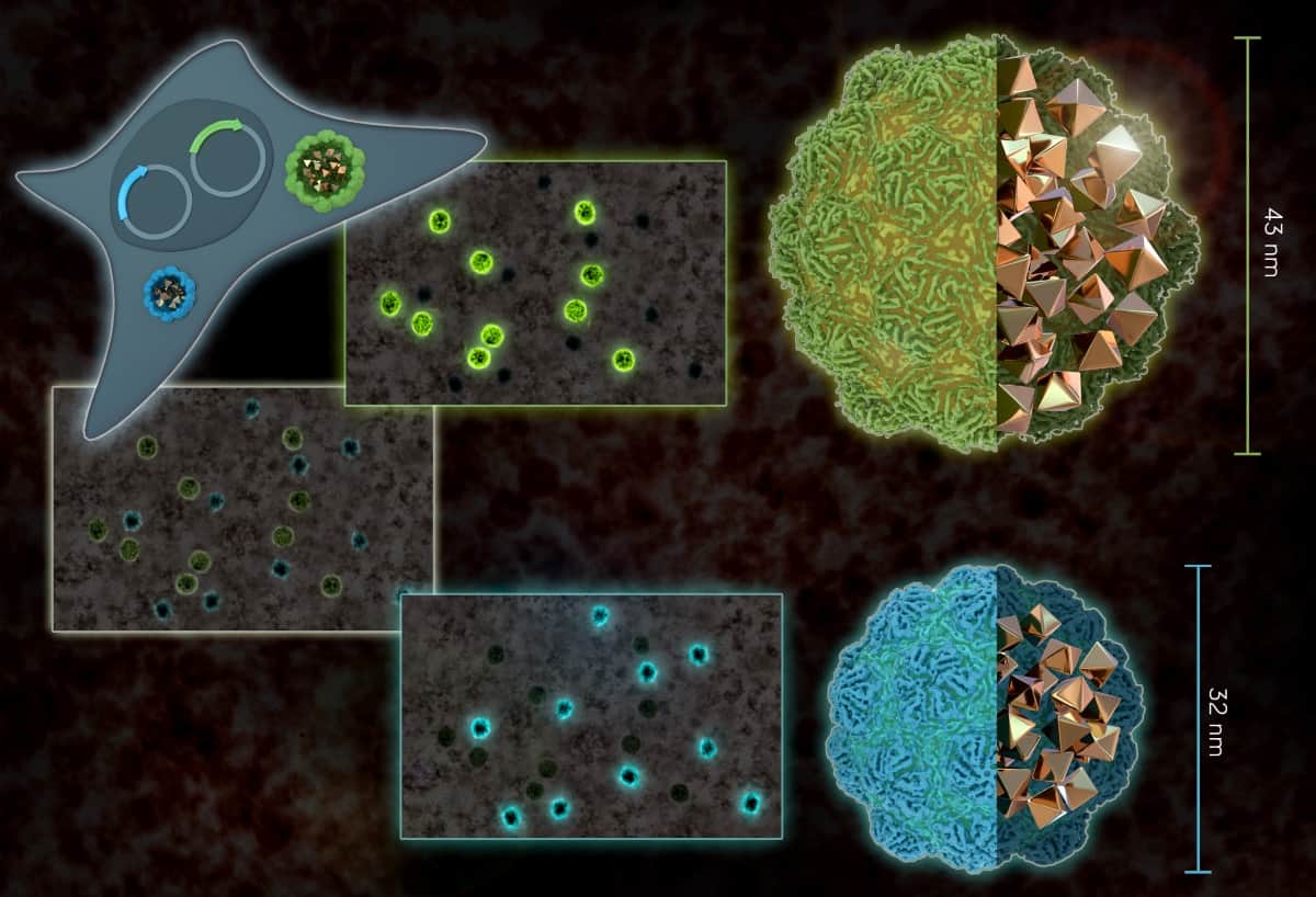
Bacterial nanostructures act as electron-microscope ...
Concatenated Metallothionein as a Clonable Gold Label for Electron ... The creation of a clonable label could provide the same advantages for electron microscopy provided that the label had high visibility in the electron microscope. Ideally, we want a small protein that would initiate formation of a heavy metal cluster from a heavy metal salt or organometallic compound. We selected the cysteine-rich protein ...
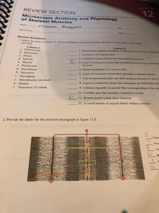
Solved 12 REVIEW SECTION Microscop of Skeletal Muscles copic ...
Ultrasensitive detection of patulin based on a Ag+-driven one … After the formation of stable Au-N bonds between AuNFs and g-C 3 N 4 nanosheets by chemical bonding, the typical transmission electron micrograph of the AuNFs/g-C 3 N 4 composite in Fig. 1 G shows that flower-like particles are fixed on a single nanosheets, indicating that AuNFs were successfully loaded onto the surface of g-C 3 N 4 nanosheets.

Transmission electron micrographs of mesophyll cells among ...
Looking at the Structure of Cells in the Microscope A typical animal cell is 10–20 μm in diameter, which is about one-fifth the size of the smallest particle visible to the naked eye. It was not until good light microscopes became available in the early part of the nineteenth century that all plant and animal tissues were discovered to be aggregates of individual cells. This discovery, proposed as the cell doctrine by Schleiden and …

AICE Biology Chapter 1: Animal Cell Electron Micrograph ...
Optical Sensor - an overview | ScienceDirect Topics 10.4.1 Sensors in cellular environments. Optical sensors operating in cellular environments can provide information about cell functions by probing molecules secreted from cells in situ and in real time without perturbing the cells. The optical properties of SWCNTs make them appealing for use in sensors under cellular environments because their emission is less likely to be absorbed …

Scanning electron micrograph of the labella of horn fly ...
Cryo‐EM reveals mechanisms of angiotensin I‐converting enzyme ... 12.07.2022 · Introduction. The zinc metalloprotease angiotensin I-converting enzyme (ACE;EC 3.4.15.1) is well-known for its crucial role in blood pressure regulation and fluid homeostasis via the renin–angiotensin–aldosterone system (RAAS), where it catalyzes the hydrolysis of angiotensin I (Ang I) to the vasopressor angiotensin II (Ang II; Ehlers & Riordan, 1989).
Proteus vulgaris bacteria, scanning electron micrograph (SEM ...
Picking faces out of a crowd: Genetic labels for identification of ... (A) Schematic of indirect (secondary) antibody labeling for correlative light and electron microscopy where the secondary antibody is conjugated to a gold bead or quantum dot. (B) Electron micrograph of microtubules (arrows) labeled with immuno-quantum dots. The primary antibody is an α-tubulin monoclonal antibody.
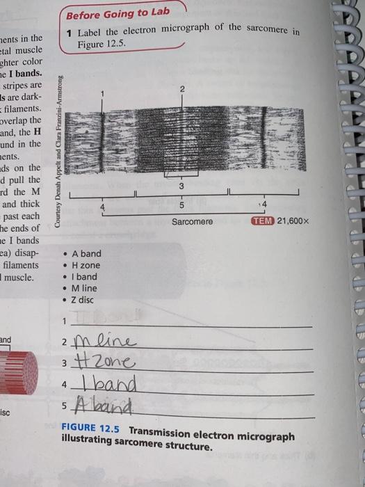
Solved Before Going to Lab Label the electron micrograph of ...
Waterproof Labels, Wet Grip, Electron Microscopy Sciences Direct thermal Labels are white and on a 1" int.Ø. core. Temperature Range: -40 to +70 °C. GRIPS to wet, frost-covered and frozen surface, Compatible with centrifuge rotors, Resistant to chemicals and solvents, Adheres to plastics, glass, metals and even paperboard boxes. Print on sheets with Laser printers or use rolls for direct thermal printers.
CcFIG 5 Legend
Label This Transmission Electron Micrograph - Kaiden Brown Label the transmission electron micrograph of the nucleus. Provide the labels for the electron micrograph in figure 12.8. Labeling for electron microscopy using antibody conjugated to. Transmission electron microscopy (tem) is a microscopy technique in which a beam of electrons is transmitted through a specimen to form an image.

B1 Muscles and Movement | Brent Cornell
Introduction to Cryo EM - Thermo Fisher Scientific - US Cryo-electron microscopy. In cryo-EM, samples are rapidly frozen (vitrified), preventing the formation of crystalline ice and preserving samples in their natural state. A transmission electron microscope (TEM) is then used to image the sample, capturing a two-dimensional view, or projection, of the specimen. By creating hundreds of projections ...

A) Scanning electron micrograph of a cross section through ...
chloroplast and mitochondrion.pdf - D SBA#: Title: Electron micrograph ... At least 2 parts labeled correctly (1 mark) 4 Annotate drawings appropriately and accurately e. 5 parts annotated correctly (4 marks)f. 4 parts annotated correctly (3 marks) g. 3 parts annotated correctly (2 marks)h.

Transmission electron micrograph of a gold-labelled Lowicryl ...
What is Electron Microscopy? - UMASS Medical School Electron microscopy (EM) is a technique for obtaining high resolution images of biological and non-biological specimens. It is used in biomedical research to investigate the detailed structure of tissues, cells, organelles and macromolecular complexes. The high resolution of EM images results from the use of electrons (which have very short ...
Solved 20 found at the beginning of the Laboratory Skeletal ...
Electron Micrographs of Cell Organelles | Zoology - Biology Discussion This is an electron-micrograph of plastid or chloroplast, which is an integral component of all green plant leaves and is characterized by following features (Fig. 15 & 16): (1) They may be spheroidal, ovoid, stellate or collar shaped and differ in size and number in different cells.

Scanning electron microscope - Wikipedia
Ch 19 Skeletal Muscle Structure Flashcards | Chegg.com

A: electron micrograph of rat neuromuscular junction treated ...

transmission electron micrograph of light cells showing ...

Rat pancreas. Electron micrograph of immunogold labeling for ...

Transmission electron micrograph of endocytosed gold-ASOR ...
What is a diagram of a plant and animal cell under an ...
Transmission electron microscope (TEM) micrograph showing the ...

The corresponds to the indicated Learning Out found at the ...
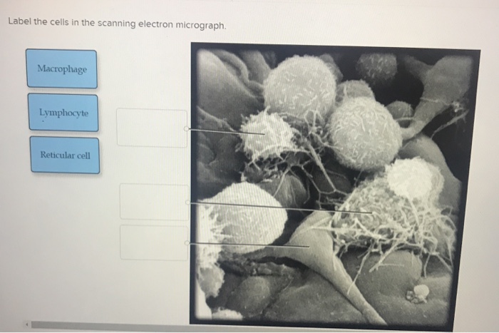
Solved Label the cells in the scanning electron micrograph ...

Electron micrograph showing a neutrophilphagocytosing ...
Post a Comment for "41 provide the labels for the electron micrograph"